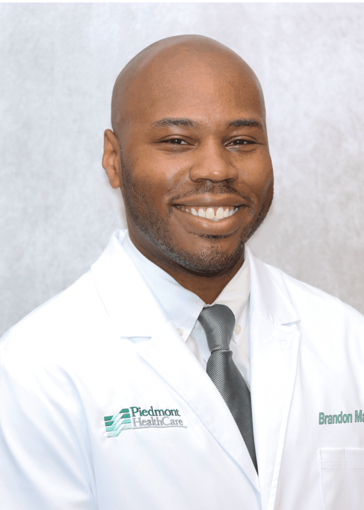
Our team is skilled in preventing, diagnosing, and treating digestive diseases using the latest medical knowledge and state-of-the-art techniques. We are dedicated to addressing the unique needs of each patient by providing the highest quality, cost-effective care in a warm, compassionate, and professional environment. Drs. Marion and Lamm are honored to offer exceptional gastroenterology and hepatology services to the residents of Iredell County and surrounding communities.
Gastroenterology
Our Approach

Our Office
2 Physicians, 1 Physician Assistant, & 1 Nurse Practitioner
Accepting New Patients
2 Convenient Locations
Statesville | Mooresville
PHC Endoscopy Center On-Site
Drs. Marion and Lamm perform procedures at PHC Endoscopy Center and Iredell Memorial Hospital
Welcome to PHC Gastroenterology!
We are dedicated to providing our patients with the best care available. Please carefully read and complete all of the patient forms prior to your appointment. We ask that you bring the completed paperwork to your visit. This will reduce your wait time once you arrive. At the time of your appointment you will need:
- All current medications or completed list
- Insurance card (s) and your co-pay
- Prescription Benefit Card if applicable
- Completed paperwork
- Copy of any applicable medical records such as lab work or procedure reports
If you are unable to keep your appointment for any reason, please contact our office as soon as possible so that we may offer your time to another patient.
Thank you for choosing PHC Gastroenterology for your gastrointestinal care! Please feel free to contact us with any questions you may have 704-878-2021.
Patient Forms
In order to speed up the check-in process from the lobby, please fill out the following New Patient Intake forms prior to your visit. Click Here to download New Patient IntakeForms
Prepare for procedures
In case you may have misplaced your paperwork from your recent visit, you can download and print your forms.
Pre-procedure Do’s and Don’ts:
Colonoscopy
Upper Endoscopy
SUPREP BOWEL PREP KIT
MOVIPREP Instructions
OSMOPREP Instructions
Golytely Prep Instructions
The Endoscopy Center
We are proud that virtually all of our gastrointestinal services are available in our Outpatient Center. The Piedmont Healthcare Endoscopy Center allows our physicians to provide the highest quality services in a friendly, comfortable, cost-efficient and convenient environment. The Endoscopy Center is equipped with the latest in medical technology. All of the physicians and staff are trained and maintain a regular quality assurance program.
We are dedicated to providing the highest quality, most efficient gastrointestinal services to our patients. Our efforts were rewarded when the Endoscopy Center became a licensed outpatient facility. That designation is awarded only after demonstrating the highest quality of care. This honor is a reflection of both our facility and staff.
Quality Assurance
Our Endoscopy Center strives to deliver the highest level of care for our patients. We achieve this by regular monitoring of specific quality measures for endoscopic performance to ensure that our Endoscopists meet the standard of care.
Our Location
Statesville
208 Old Mocksville Road
Statesville, NC 28677
Phone 704-878-2021
Mooresville
359 Williamson Road,
Mooresville, NC 28117
Phone 704-235-1829
Call today to schedule
an appointment
Conditions
Procedures
Upper Endoscopy (EGD)
Upper endoscopy, also called esophagogastroduodenoscopy, or EGD, uses a thin scope with a light and camera at its tip to look inside of the upper digestive tract — the esophagus, stomach, and first part of the small intestine, called the duodenum.
Usually performed as an outpatient procedure, upper endoscopy sometimes must be performed in the hospital or emergency room to both identify and treat conditions such as upper digestive system bleeding.
The procedure is commonly used to help identify the causes of:
- Abdominal or chest pain
- Nausea and vomiting
- Heartburn
- Bleeding
- Swallowing problems
Capsule Endoscopy “Camera Pill”
What is Capsule Endoscopy?
Capsule Endoscopy lets your doctor examine the lining of the middle part of your gastrointestinal tract, which includes the three portions of the small intestine (duodenum, jejunum, ileum). Your doctor will use a pill sized video capsule called an endoscope, which has its own lens and light source and will view the images on a video monitor. You might hear your doctor or other medical staff refer to capsule endoscopy as small bowel endoscopy, capsule enteroscopy, or wireless endoscopy.
Why is Capsule Endoscopy Done?
Capsule endoscopy helps your doctor evaluate the small intestine. This part of the bowel cannot be reached by traditional upper endoscopy or by colonoscopy. The most common reason for doing capsule endoscopy is to search for a cause of bleeding from the small intestine. It may also be useful for detecting polyps, inflammatory bowel disease (Crohn’s disease), ulcers, and tumors of the small intestine.
As is the case with most new diagnostic procedures, not all insurance companies are currently reimbursing for this procedure. You may need to check with your own insurance company to ensure that this is a covered benefit.
How Should I Prepare for the Procedure?
An empty stomach allows for the best and safest examination, so you should have nothing to eat or drink, including water, for approximately twelve hours before the examination. Your doctor will tell you when to start fasting.
Tell your doctor in advance about any medications you take including iron, aspirin, bismuth subsalicylate products and other “over-the-counter” medications. You might need to adjust your usual dose prior to the examination. Discuss any allergies to medications as well as medical conditions, such as swallowing disorders and heart or lung disease. Tell your doctor of the presence of a pacemaker, previous abdominal surgery, or previous history of obstructions in the bowel, inflammatory bowel disease, or adhesions.
What Can I Expect During Capsule Endoscopy?
Your doctor will prepare you for the examination by applying a sensor device to your abdomen with adhesive sleeves (similar to tape). The capsule endoscope is swallowed and passes naturally through your digestive tract while transmitting video images to a data recorder worn on your belt for approximately eight hours. At the end of the procedure you will return to the office and the data recorder is removed so that images of your small bowel can be put on a computer screen for physician review.
Most patients consider the test comfortable. The capsule endoscope is about the size of a large pill. After ingesting the capsule and until it is excreted, you should not be near an MRI device or schedule an MRI examination.
ERCP-Diagnostic and Therapeutic
An endoscopic retrograde cholangiopancreatogram (ERCP) is a test that combines the use of a flexible, lighted scope (endoscope) with X-ray pictures to examine the tubes that drain the liver, gallbladder, and pancreas.
The endoscope is inserted through the mouth and gently moved down the throat into the esophagus, stomach, and duodenum until it reaches the point where the ducts from the pancreas (pancreatic ducts) and gallbladder (bile ducts) drain into the duodenum.
ERCP can treat certain problems found during the test. If an abnormal growth is seen, an instrument can be inserted through the endoscope to obtain a sample of the tissue for further testing (biopsy).
Why It Is Done
ERCP is done to:
- Check persistent abdominal pain or jaundice.
- Find gallstones or diseases of the liver, bile ducts, or pancreas.
- Remove gallstones from the common bile duct if they are causing a problem such as blockage (obstruction), inflammation or infection of the common bile duct (cholangitis), or pancreatitis.
- Open a narrowed bile duct or insert a drain.
- Get a tissue sample for further testing (biopsy)
Hydrogen Breath Testing
The hydrogen breath test is a test that uses the measurement of hydrogen in the breath to diagnose several conditions that cause gastrointestinal symptoms.
How is hydrogen breath testing performed?
Prior to hydrogen breath testing, the patient fasts for at least 12 hours. At the start of the test, the patient blows into and fills a balloon with a breath of air. The concentration of hydrogen is measured in a sample of breath removed from the balloon. The patient then ingests a small amount of the test sugar (lactose, sucrose, sorbitol, fructose, lactulose, etc. depending on the purpose of the test).
Additional samples of breath are collected and analyzed for hydrogen every 15 minutes for three and up to five hours.
H Pylori Testing
Helicobacter pylori tests are used to detect a Helicobacter pylori (H. pylori) infection in the stomach and upper part of the small intestine (duodenum). H. pylori can cause peptic ulcers. But most people with H. pylori in their digestive systems do not develop ulcers.
Four tests are used to detect H. pylori:
Blood antibody test. A blood test checks to see whether your body has made antibodies to H. pylori bacteria. If you have antibodies to H. pylori in your blood, it means you either are currently infected or have been infected in the past.
Urea breath test. A urea breath test checks to see if you have H. pylori bacteria in your stomach. This test can show if you have an H. pylori infection. It can also be used to see if treatment has worked to get rid of H. pylori. The breath test is not always available.
Stool antigen test. A stool antigen test checks to see if substances that trigger the immune system to fight an H. pylori infection (H. pyloriantigens) are present in your feces (stool). Stool antigen testing may be done to help support a diagnosis of H. pylori infection or to determine whether treatment for an H. pylori infection has been successful.
Stomach biopsy. A small sample (biopsy) is taken from the lining of your stomach and small intestine during an endoscopy. Several different tests may be done on the biopsy sample. For more information, see the medical test Upper Gastrointestinal Endoscopy.
Why It Is Done
A Helicobacter pylori (H. pylori) test is done to:
- Determine whether an infection with H. pylori bacteria may be causing an ulcer or irritation of the stomach lining (gastritis).
- Determine whether treatment for an H. pylori infection has been successful.
Hemorrhoid Removal
For those of you that have bothersome hemorrhoids after using conservative measures such as topical creams, ointments and warm sitz baths, you may want to consider a minimally invasive procedure.
Rubber band ligation (hemorrhoid banding) is the most widely used procedure. It is painless, quick (<1min) and successful in approximately 80 percent of patients.
Rubber bands or rings are placed around the base of an internal hemorrhoid. As the blood supply is restricted, the hemorrhoid shrinks and degenerates over several days. Many patients report a sense of “tightness” after the procedure, which may improve with warm sitz baths.
Patients are encouraged to use fiber supplements to avoid constipation. Three banding sessions are performed every two weeks for removal of all three hemorrhoid columns.
Colon Cancer Screening
What is colonoscopy?
Colonoscopy is a procedure used to see inside the colon and rectum. Colonoscopy can detect inflamed tissue, ulcers, and abnormal growths. The procedure is used to look for early signs of colorectal cancer and can help doctors diagnose unexplained changes in bowel habits, abdominal pain, bleeding from the anus, and weight loss.
What are the colon and rectum?
The colon and rectum are the two main parts of the large intestine. Although the colon is only one part of the large intestine, because most of the large intestine consists of colon, the two terms are often used interchangeably. The large intestine is also sometimes called the large bowel.
The colon and rectum are the two main parts of the large intestine.
Digestive waste enters the colon from the small intestine as a semisolid. As waste moves toward the anus, the colon removes moisture and forms stool. The rectum is about 6 inches long and connects the colon to the anus. Stool leaves the body through the anus. Muscles and nerves in the rectum and anus control bowel movements.
How is colonoscopy performed?
Examination of the Large Intestine
During colonoscopy, patients lie on their left side on an examination table. In most cases, a light sedative, and possibly pain medication, helps keep patients relaxed. Deeper sedation may be required in some cases. The doctor and medical staff monitor vital signs and attempt to make patients as comfortable as possible.
During colonoscopy, patients lie on their left side on an examination table.
The doctor inserts a long, flexible, lighted tube called a colonoscope, or scope, into the anus and slowly guides it through the rectum and into the colon. The scope inflates the large intestine with carbon dioxide gas to give the doctor a better view. A small camera mounted on the scope transmits a video image from inside the large intestine to a computer screen, allowing the doctor to carefully examine the intestinal lining. The doctor may ask the patient to move periodically so the scope can be adjusted for better viewing. Once the scope has reached the opening to the small intestine, it is slowly withdrawn and the lining of the large intestine is carefully examined again. Bleeding and puncture of the large intestine are possible but uncommon complications of colonoscopy.
Removal of Polyps and Biopsy
A doctor can remove growths, called polyps, during colonoscopy and later test them in a laboratory for signs of cancer. Polyps are common in adults and are usually harmless. However, most colorectal cancer begins as a polyp, so removing polyps early is an effective way to prevent cancer.
The doctor can also take samples from abnormal-looking tissues during colonoscopy. The procedure, called a biopsy, allows the doctor to later look at the tissue with a microscope for signs of disease.
The doctor removes polyps and takes biopsy tissue using tiny tools passed through the scope. If bleeding occurs, the doctor can usually stop it with an electrical probe or special medications passed through the scope. Tissue removal and the treatments to stop bleeding are usually painless.
Recovery
Colonoscopy usually takes 30 to 60 minutes. Cramping or bloating may occur during the first hour after the procedure. The sedative takes time to completely wear off. Patients may need to remain at the clinic for 1 to 2 hours after the procedure. Full recovery is expected by the next day. Discharge instructions should be carefully read and followed.
Patients who develop any of these rare side effects should contact their doctor immediately:
- severe abdominal pain
- fever
- bloody bowel movements
- dizziness
- weakness
At what age should routine colonoscopy begin?
Routine colonoscopy to look for early signs of cancer should begin at age 50 for most people. If there is a family history of colorectal cancer, a personal history of inflammatory bowel disease, or other risk factors, checks should be performed earlier. The doctor can advise patients about how often to get a colonoscopy.
Points to Remember
- Colonoscopy is a procedure used to see inside the colon and rectum.
- All solids must be emptied from the gastrointestinal tract by following a clear liquid diet for 1 to 3 days before colonoscopy.
- During colonoscopy, a sedative, and possibly pain medication, helps keep patients relaxed.
- A doctor can remove polyps and biopsy abnormal-looking tissues during colonoscopy.
- Driving is not permitted for 12 hours after colonoscopy to allow the sedative time to wear off.


